Lung Cancer
INTRODUCTION
Lung cancer occurs when cells in the lung grow abnormally out of control and form tumors. Lung cancer is the leading cause of cancer-related death in the United States. Smoking causes the majority of lung cancer cases. Early stages of lung cancer may not produce symptoms; however, early diagnosis and treatment is important to ensure the best outcome. Lung cancer may be treated with surgery, radiation, and chemotherapy.
ANATOMY
Your lungs are located inside the ribcage in your chest. Your diaphragm is beneath your lungs. The diaphragm is a dome-shaped muscle that works with your lungs when you breathe.
From your nose and mouth, air travels towards your lungs through a series of tubes. The trachea or windpipe is located in your throat. The bottom of the trachea separates into two large tubes called the main stem bronchi. The left main stem bronchus goes into the left lung, and the right main stem bronchus goes into the right lung.
Inside the lung, the bronchi branch off and become smaller. These smaller air tubes are called bronchioles. There are approximately 30,000 bronchioles in each lung. The end of each bronchiole has tiny air sacs called alveoli. There are about 600 million alveoli in your lungs. Each alveolus is covered in small blood vessels called capillaries. The capillaries move oxygen in and carbon dioxide out of your blood.
When you breathe air in or inhale, your diaphragm flattens and your ribs relax outward to allow your lungs to expand. The air that you inhale through your nose or mouth travels down the trachea. Tiny hair-like structures in the trachea, called cilia, filter the air to help keep mucus and dirt out of your lungs. The air travels through the bronchi and the bronchioles and into the alveoli. Oxygen in the air passes through the alveoli into the capillaries. The oxygen attaches to red blood cells and travels to the heart. The heart sends the oxygenated blood to the cells in your body.
When you breathe air out or exhale, the process is the opposite of when you inhale. Once your body has used the oxygen in the blood, the deoxygenated blood returns to the capillaries. The blood now contains carbon dioxide and waste products that must be removed from your body. The capillaries transfer the carbon dioxide and wastes from the blood and into the alveoli. The air travels through the bronchioles, the bronchi, and the trachea. As you exhale, your diaphragm rises and your ribs move inward. As your lungs compress, the air is released out of your mouth or nose.
CAUSES
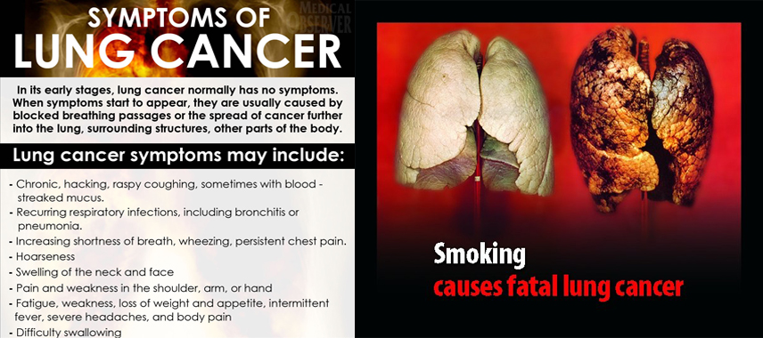
Smoking is the most common cause of lung cancer. Cancer occurs when cells grow abnormally and out of control, instead of dividing in an orderly manner. Exposure to second-hand smoke, polluted air or water, and certain industrial chemicals increases the risk for lung cancer. However, people that have never smoked may also develop lung cancer.
There are different types of lung cancer that are categorized as non-small cell lung cancer (NSCLC) or small cell lung cancer (SCLC). NSCLC is the most common type. NSCLC tends to grow and spread slowly. Three forms of NSCLC are:
Adenocarcinomas- NSCLC
Adenocarcinomas are generally located in the outer portion of the lung.
Squamous Cell Carcinomas- NSCLC
Squamous cell carcinomas generally originate in the center of the lung by a bronchus.
Large Cell Carcinomas- NSCLC
Large cell carcinomas can occur in any part of the lung. They are the fastest growing and spreading type of NSCLC
Small Cell Lung Cancer (SCLC)
SCLC is the fastest growing type of lung cancer. SCLC can be very aggressive and spread quickly to other parts of the body, such as the brain, liver, and bones. SCLC can cause large tumors. The most common type of SCLC is oat cell cancer, which is named for the shape of the cancer cells. SCLC may also be categorized as mixed small cell/large cell carcinoma or combined small cell carcinoma. Smoking is almost always the cause of SCLC.
SYMPTOMS

Lung cancer may not cause symptoms or may cause only minimal symptoms in the early stages. Symptoms of lung cancer include a new cough that will not go away or worsening of a chronic cough, such as a smoker’s cough. You may cough up blood and/or develop pneumonia and bronchitis repeatedly. You may experience changes in the way you breathe, such as wheezing and shortness of breath. You may have chest pain. You may lose your appetite and lose weight without trying. You may feel tired all of the time. Less common signs include swallowing problems, a hoarse voice, back pain, joint pain, weakness, and facial swelling, drooping, or paralysis.
DIAGNOSIS
Your doctor can begin to diagnose lung cancer by reviewing your medical history and conducting a physical examination and some tests. You should tell your doctor if you smoke or if you have ever smoked. Your doctor will listen to your lungs with a stethoscope while you breathe. You may perform breathing or pulmonary tests to assess how your lungs are functioning. Your sputum may be tested for infection.
Chest X-rays, magnetic resonance imaging (MRI) scans, and computerized tomography (CT) scans are used to provide images of your lungs to help diagnose cancer. A bronchoscopy may be used to view the inside of your lungs. This test involves inserting a thin fiberoptic tube through your windpipe (trachea) and into your lungs. A thoracoscopy is used to examine the lining inside and outside of your lungs. A biopsy may be taken of tissue or fluids to examine the cells for cancer. A biopsy can confirm the diagnosis and identify the type of lung cancer.
If you have lung cancer, your doctor will assign your cancer a classification stage based on the results of all of your tests. Staging describes the cancer and how it has metastasized. Cancer that has spread from its original site to other parts of the body is termed metastatic cancer. Staging is helpful for treatment planning and recovery prediction.
There is more than one type of staging system for cancer, and you should make sure that you and your doctor are referring to the same one. Generally, lower numbers in a classification system indicate a less serious cancer, and higher numbers indicate a more serious cancer. The stages may be subdivided into classifications that use letters and numbers.
STAGES OF NON-SMALL CELL LUNG CANCER
Stage I: The cancer is located only in the lungs and has not spread to any lymph nodes.
Stage II: The cancer is in the lung and nearby lymph nodes.
Stage III: Cancer is found in the lung and in the lymph nodes in the middle of the chest, also described as locally advanced disease. Stage III has two subtypes:
• If the cancer has spread only to lymph nodes on the same side of the chest where the cancer started, it is called stage IIIA.
• If the cancer has spread to the lymph nodes on the opposite side of the chest, or above the collar bone, it is called stage IIIB.
Stage IV: This is the most advanced stage of lung cancer, and is also described as advanced disease. This is when the cancer has spread to both lungs, to fluid in the area around the lungs, or to another part of the body, such as the liver or other organs.
TREATMENT
The type of treatment that you receive depends on several factors, including the type of lung cancer that you have. Treatment may be used to reduce the size of a tumor, prevent the cancer from spreading, or cure the cancer. If the cancer cannot be cured, treatments may be used to reduce the symptoms and prolong life.
Small cell lung cancer responds well to radiation therapy and chemotherapy. Non-small cell lung cancer may be treated with surgery to remove the cancer, part of the lung, or all of a lung. Surgery may be followed by chemotherapy or radiation therapy. Chemotherapy involves using a series of cancer-fighting drugs over a period of time. There are several types of radiation therapies for lung cancer.
Radiation therapy is a painless procedure. Your doctor will prescribe the amount and length of treatment based on your condition. Radiation therapy is usually delivered in several doses over a period of time. There are different types of radiation therapy that may be used to help treat lung cancer.
External beam radiation uses high-energy beams to disrupt the growth of cancer cells. Radiation damages all cells both healthy and cancerous in the exposed area. Radiated cancer cells are not able to repair themselves or replicate. Your doctor may prescribe medication to prevent the side effects of radiation.
State-of-the-art technology has advanced radiation methods to help make them more effective and tolerable. Three dimensional conformal radiation therapy (3D CRT) maps a tumor with imaging scans before treatment. Magnetic resonance imaging (MRI) scans, computed tomography (CT) scans, and positron emission tomography (PET) scans are used to produce images of a tumor and its surrounding tissue. The images from the scans are combined with a computer called a multi-leaf collimator (MLC). The MLC produces a 3D image of the cancer and formulates a treatment plan specifically for the 3D image. Four dimensional conformal radiation therapy (4D CRT) is able to adjust the radiation for the movements of breathing. It is useful for treating lung cancers. This allows direct multiple beams of radiation to be targeted precisely at the tumor and spare as much healthy tissue as possible.
Intensity modulated radiation therapy (IMRT) is a refined type of 3D CRT. IMRT allows the radiologist to sculpt the edges of a tumor, sparing healthy tissue. With IMRT, the radiation dose can be changed during a treatment session. In the past, radiation was delivered in one dose, from the beginning to the end of a treatment session. IMRT allows the radiation dose to alter and conform more specifically to the shape of the tumor while minimizing the dose received by healthy tissues.
Image-Guided Radiation Therapy (IGRT) is a type of external beam radiation therapy for cancer that adjusts for the size and shape of a cancer tumor throughout treatment. Cancer tumors may move, change size, and change shape throughout the course of treatment. IGRT uses advanced imaging technology to visualize the tumor before each treatment. Based on the daily images, the radiation is configured before each treatment. In turn, the radiation is more precise, effective, and results in fewer side effects.
Stereotactic Radiosurgery (SRS) and Stereotactic Radiotherapy (SRT) are both methods of delivering radiation therapy to lung cancer tumors. SRS or SRT may be used instead of or along with surgery. SRS involves a single radiation treatment. SRT uses a series of treatments over time. Both methods spare healthy tissues because the radiation precisely targets the cancer.
With SRS and SRT, sophisticated software controls the radiation treatment beams to match the exact shape of a tumor or lesion. The beams may be moved to penetrate the cancer from different angles. The state-of-the-art equipment is able to adjust for minor patient movements. This precision delivery method quickly allows the cancer to receive the full dose of radiation, while the surrounding healthy tissue only receives a small percentage of radiation.
The experience of cancer and cancer treatments can be a very emotional experience for you and your loved ones. It is important to embrace positive sources of support. Some people find comfort in their families, friends, co-workers, counselors, and faith. Cancer support groups are a helpful resource where you can receive support, information, and understanding from people with similar experiences. Ask your doctor for support groups near you.
PREVENTION

Smoking causes most cases of lung cancer. You may prevent cancer by not smoking or quit smoking. If it is difficult for you to quit smoking on your own, ask your doctor about medications and resources to help you. You should avoid exposure to second-hand smoke.
You should have your home or business checked for radon gas. Radon gas rises from the soil beneath a home and may enter it through cracked foundation, pipes, or other openings. You cannot see or smell radon, and the only way to know if it is present is to have a specific test for it. Radon gas levels may be corrected if they are high.
Make sure that you are following safe working practices if you are exposed to asbestos, uranium, arsenic, and certain petroleum products at your job. Workers that smoke and are exposed to such chemicals have a greater risk of developing lung cancer.
AM I AT RISK
Risk factors may increase your likelihood of developing lung cancer, although some people that develop lung cancer do not have any risk factors. People with all of the risk factors may never develop the disease; however, the chance of developing lung cancer increases with the more risk factors you have. You should tell your doctor about your risk factors and discuss your concerns.
RISK FACTORS FOR LUNG CANCER:
Cigarette smoking causes the majority of lung cancer. Smoking pipe tobacco or cigars also can contribute to lung cancer. However, people that have never smoked may develop lung cancer.
Inhaling second-hand smoke from others increases your risk of lung cancer.
Exposure to high levels of air pollution increases the risk of lung cancer.
High levels of arsenic in drinking water are associated with lung cancer.
Radon gas in homes or businesses can cause lung cancer.
You have a greater risk for lung cancer if your family members have had lung cancer.
Lung scarring from chronic emphysema, bronchitis, and other conditions can raise the risk of lung cancer.
Exposure at your job to cancer-causing substances, such as asbestos, uranium, arsenic, and certain petroleum products, increases the risk of developing lung cancer. People that are exposed to such products and smoke have even a greater risk.
Radiation therapy to the lungs can increase the risk of lung cancer.
COMPLICATIONS
Lung cancer can spread to other parts of the body, including the other lung, brain, bones, lymph nodes, and liver. Some types of lung cancer are aggressive and can spread quickly. Lung cancer may recur or come back after it has been treated. Lung cancer is the leading cause of cancer-related death in the United States.
References:
http://en.wikipedia.org/wiki/Lung_cancer
http://www.lungcancer.org/


